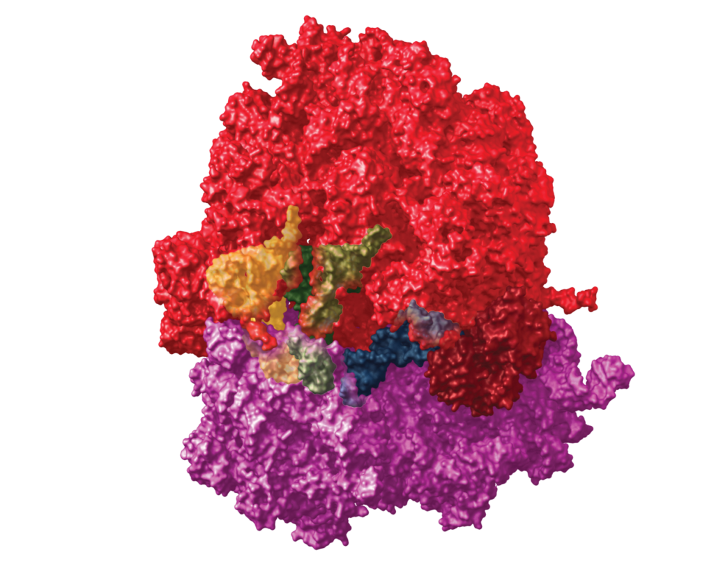
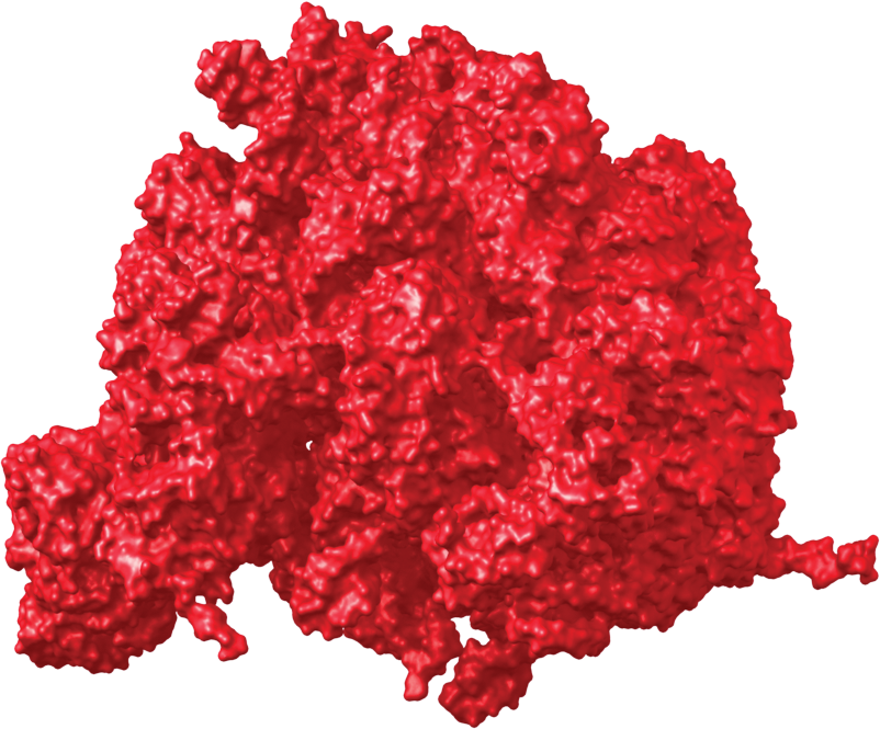
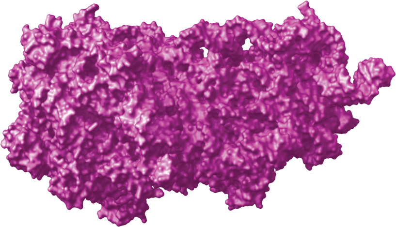
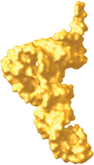
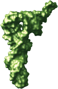
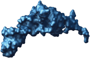
This illustration combined two PDB files, 1GIX and 1GIY. Within Chimera, I was able to isolate the 50S and 30S sections of the ribosome and export them separately. The same method was used to export the E, P, and A sites.
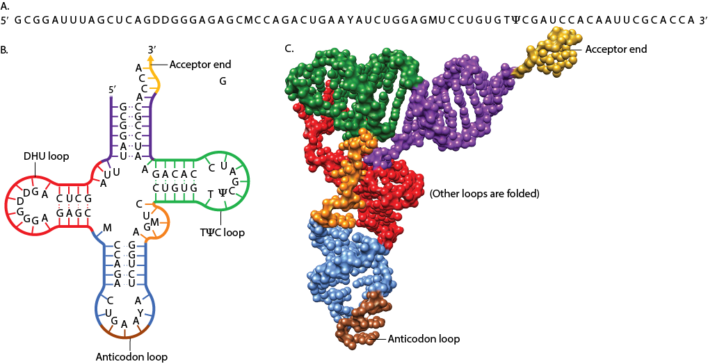
tRNA structure
This illustration show 3 different representations of tRNA. (1GIX). The original molecular structure is a single image. With Chimera I was able to isolate the loops and color them to coincide with the vector loops.
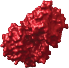
RNA polymerase
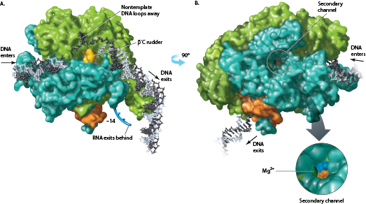
This 3D structure was rendered from the PDB file 1L9Z. Because it is 3D, I was able to render part A, rotate the structure 90 degrees, and then render that view. I was also able to isolate the magnesium ion in the secondary channel so I could create the enlargement.
